Revolutionizing MRI Technology with PHIP+
How Does Our PHIP+ Process Work?
In scientific terms, we have developed a precursor molecule, a pre-stage of the metabolite, that contains an unsaturated side arm in addition to the metabolite pyruvate. This precursor can be hydrogenated with inexpensive parahydrogen at the side arm, and the resulting high spin order of the transferred hydrogen protons can be used to create hyperpolarization in the ¹³C nucleus of pyruvate through a targeted spin order transfer. The relatively long lifetime of the hyperpolarization in this ¹³C nucleus then allows the side arm to be cleaved off, isolating the hyperpolarized pyruvate for injection.
But how exactly does our PHIP+ process work? How can we hyperpolarize endogenous metabolites?
If you're not a physicist or chemist, many of these terms may not be familiar to you. So here's a more detailed explanation to help you better understand the advantages of our process and, in particular, our patented metabolic tracer.
Some atomic nuclei, such as hydrogen nuclei or ¹³C nuclei, behave like tiny magnets in a magnetic field and align themselves accordingly. In MRI, the behavior of the overall "magnetization" generated by all these atomic mini-magnets is used to create an image. (Technically, it is the induced electric current that is measured.)
The issue is that almost half of these atomic magnets behave exactly opposite to the other half, causing their magnetic signals to almost completely cancel each other out. A small fraction remains uncanceled, creating a measurable signal. Only this weak signal from the excess magnetization contributes to the final MRI image. This excess magnetization is also often referred to as polarization.
However, the number of atoms that contribute to this excess magnetization (or polarization) is extremely low in conventional MRI. In a typical MRI scan, only about 1 in 1 million atomic nuclei contributes to the measurable signal, while the remaining 999,999 nuclei are invisible to the MRI. This is why only very abundant atomic nuclei, such as protons (since the human body is composed of about 80% water, with each water molecule containing two protons), can generate a signal strong enough to be detected in a reasonable timeframe.
To deal with this weak signal in conventional MRI, there are two main strategies:
- Increase the magnetic field strength – This is extremely expensive and has physical limitations. To achieve the same signal strength as hyperpolarized MRI with conventional MRI alone, magnetic field strengths comparable to those of a neutron star would be required, which remains firmly in the realm of science fiction.
- Repeat the measurement multiple times and sum the signals – This improves the signal-to-noise ratio but requires taking multiple images and creating an overlay, which increases scan time. Moreover, fast and dynamic processes in the body, such as metabolism, can never be detected using this approach, because each image would differ from the previous one, making an overlay impossible.
To illustrate the sensitivity problem in conventional MRI, imagine that the excess magnetization is represented by water in a 1-liter beaker. If the beaker is empty, all spins cancel each other out completely, resulting in no measurable MRI signal. If the beaker is completely full, all mini-magnets align in the same direction, and their combined magnetic fields produce the strongest possible MRI signal.
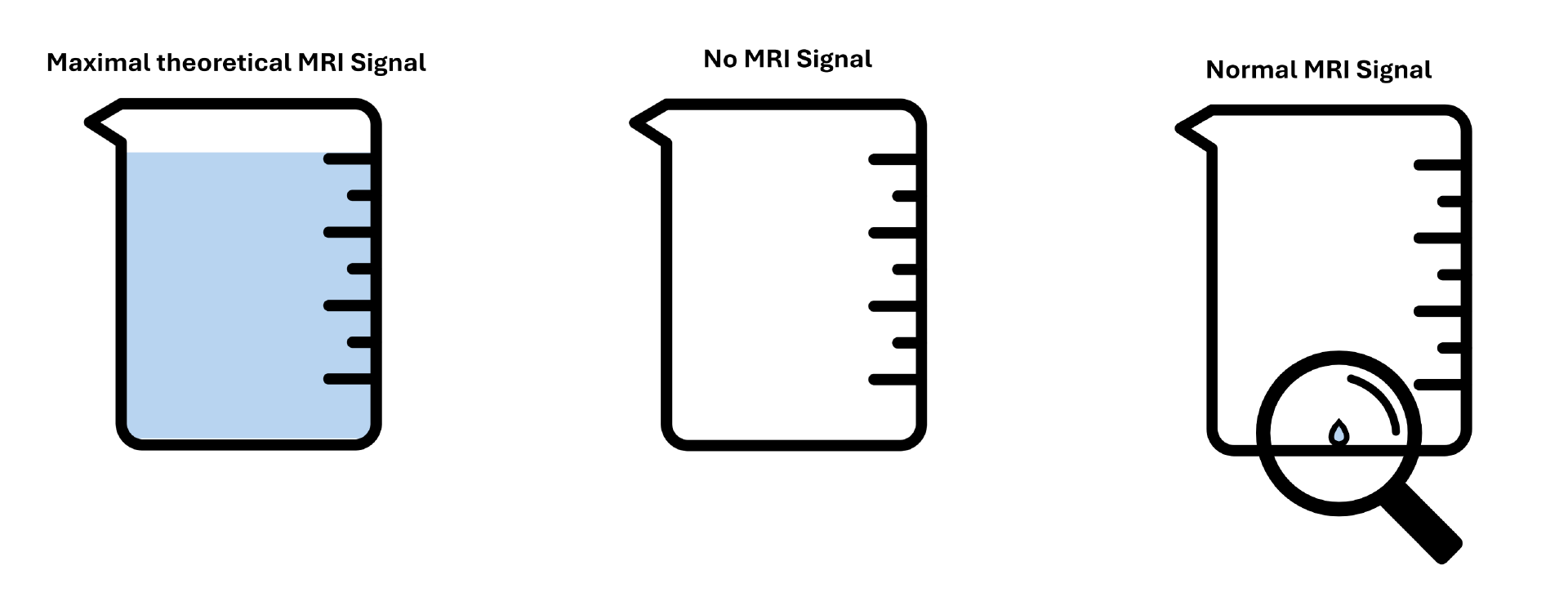
In standard MRI, however, the beaker is barely filled, containing only about one microliter of water. Yes, you read that correctly: not a milliliter, but a microliter, roughly 1,000 times smaller than a droplet. This tiny signal is what clinical MRI relies on to generate black-and-white images of your organs. Because the signal is so weak, only the most abundant atoms in the human body, hydrogen nuclei, can be detected. These include hydrogen atoms in water, fats, carbohydrates, and proteins, which make up different tissue types.
As mentioned earlier, multiple images must be acquired and combined to produce the final image seen in clinical MRI. This is one reason why some MRI scans take a long time, requiring patients to remain still, as any movement between scans can lead to image blurring.
What Does Hyperpolarization Do?
In simple terms, hyperpolarization fills the beaker beyond the tiny droplet, meaning it amplifies the measurable MRI signal.
Sounds simple, right? In reality, it’s quite complex.
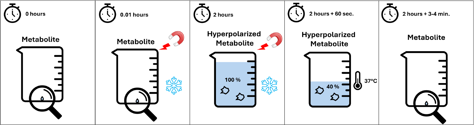
If we were to directly fill the beaker (increase the measurable MRI signal) of our tracer, extreme conditions would typically be required. The most widely used clinical research method, dDNP (dissolution Dynamic Nuclear Polarization), relies on extremely strong magnetic fields and ultra-low temperatures near absolute zero (very cold and very expensive) to hyperpolarize small amounts of a metabolite just before measurement. This method is cumbersome, costly, and time-consuming, requiring several hours of preparation for each measurement.
Another challenge is that if we fill the beaker beyond the normal microliter level, it quickly “leaks.” Within 3 to 5 minutes, the extra signal is lost, returning to its original weak state. In physics, this is known as relaxation, where the system naturally returns to equilibrium (an almost empty beaker with almost no signal intensity).
At first glance, this may seem like a bad deal: a highly complex process that significantly enhances the MRI signal but is expensive and short-lived.
We Do It Better
However, we use clever chemical and physical tricks to achieve high signal enhancement quickly and affordably, while ensuring the hyperpolarized tracer remains detectable long enough for a high-quality MRI scan.
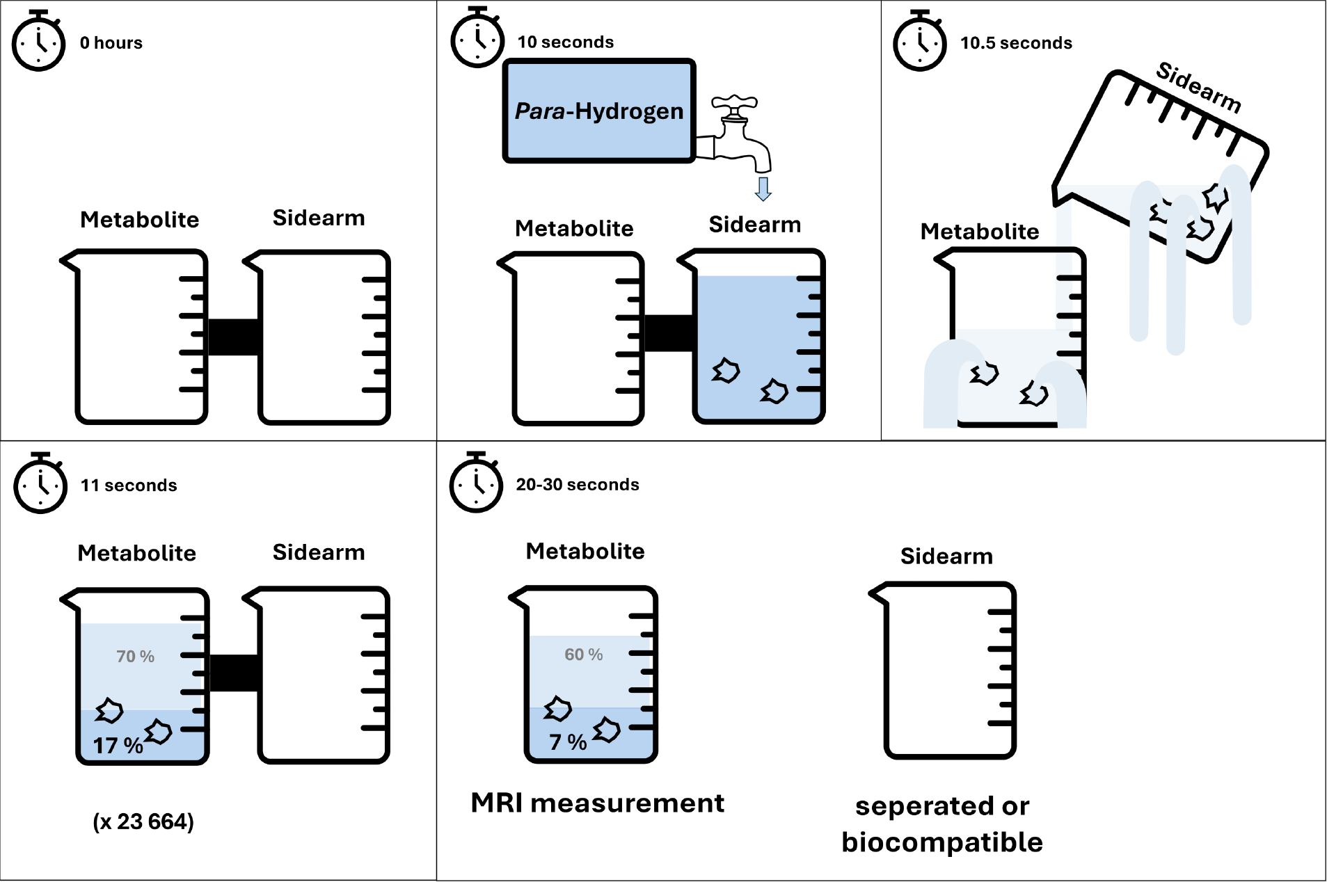
First, instead of hyperpolarizing the metabolite directly, we use an unsaturated side arm. This side arm also has its own 1-liter beaker.
We hydrogenate this side arm with parahydrogen, a special quantum form of hydrogen. This is equivalent to filling the side arm's beaker under a running tap until it's full. The huge advantage of this approach is that it is incredibly fast (seconds!), uses cheap and widely available parahydrogen, and parahydrogen can be stored for months. Essentially, parahydrogen serves as a perfect water reservoir for us (in reality, a highly accessible and cost-effective source of spin order).
After filling the side arm’s beaker, we need to act quickly because this beaker is also very leaky. We transfer the water from the side arm’s beaker to the tracer’s beaker. Unfortunately, some water is lost during the transfer, and both beakers continue to leak.
Our competitors, who use a similar approach with a different tracer, typically achieve a fill level of around 200 mL in the tracer’s beaker. That’s impressive, but we can do better.
With our patented metabolic tracer, which is isotope-labeled in both the metabolite and the side arm, we reduce the number of leaks in both beakers. Additionally, we use a specially designed vinyl side arm, which shortens the transfer distance and reduces spillage.
As a result, we have successfully demonstrated higher signal retention. Our improvements have already enabled us to reach fill levels of around 300 ml in the tracer's beaker. Calculations suggest that by speeding up and further automating the transfer process, we could achieve 600–700 ml.
Furthermore, about one-third of the leaks in the tracer’s beaker have been sealed, allowing the hyperpolarization to last longer and enabling higher signal strength and extended measurement times.