Redefining Cancer Diagnostics and Treatment with Metabolic Imaging
Explanation: Metabolic MRI
A picture says more than a thousand words – this is what makes hyperpolarization possible!
Scientific concepts are often highly complex, and the underlying details can be difficult to understand. Metaphors help make these concepts more tangible and convey their meaning in an intuitive way. This is also true for hyperpolarization in MRI and metabolic imaging, whose mechanisms are difficult to grasp without illustrative comparisons. Here is our attempt to explain hyperpolarization and metabolic MRI in an understandable way.
Hyperpolarization
Imagine you want to take a picture in a dark room. A conventional MRI scan is like taking a photo in poor lighting. The image is grainy, and details are difficult to recognize. Conventional approaches to signal enhancement using even stronger MRI magnets work, but they are like trying to brighten the photo by installing large, expensive floodlights. This is not only technically challenging but also very expensive. Just think about the cost of purchasing multiple floodlights and the electricity required to power them.
Another problem is that the structures you want to capture in the image only appear in different shades of gray, and some are almost transparent. Simply adding more light will eventually no longer improve the image. Additionally, you will likely never be able to clearly depict transparent structures.
Our approach works completely differently: instead of making the entire image brighter, hyperpolarization selectively enhances specific objects in the image. It is as if the structures you want to capture in your image suddenly light up in color. This not only provides a clearer representation in an otherwise dark room but also allows nearly completely transparent structures (diluted metabolite) to be seen in the image that were previously hidden.
It is important to mention that these substances do not actually "glow." In general, MRI and hyperpolarization are techniques that do not involve harmful radiation, such as radioactivity. The "glow" corresponds to the magnetic alignment of atomic nuclei. This is a completely harmless physical state that disappears after a very short time and has no effects on the body.
Metabolic MRI – Visualizing Metabolism in Real Time
While classical MRI can only depict tissue, hyperpolarized MRI makes it possible to make selected metabolites appear to "glow" with very high intensity in the MRI image. The effect of this "glow" goes even further: when the molecule is absorbed and metabolized by the body, the glow is temporarily transferred to the resulting metabolic products. These products also become visible in the MRI and can even be distinguished from each other based on the "color" of their glow. We can now not only see where a substance is being metabolized but also determine into which products it is being broken down and in what ratio they are produced at each location in the human body.
Due to the high signal amplification of our technique, it is possible to visualize metabolism in real time using MRI. This is a groundbreaking development that was previously not possible in such a simple and practical way. But why is this so important for diagnostics and treatment?
The Warburg Effect – Known for Over 100 Years, but Previously Invisible
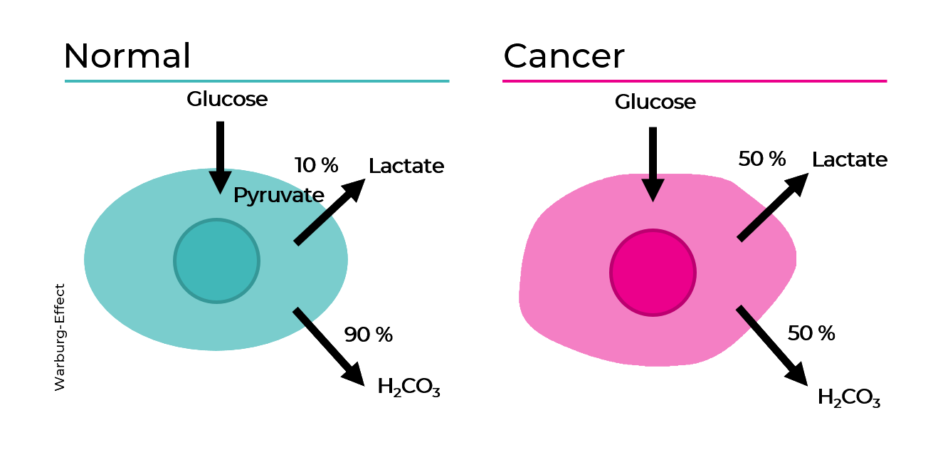
The metabolism of a healthy cell follows clear rules. However, in some diseases, the metabolism changes at a cellular level. The most well-known example of a disease that alters cellular metabolism is cancer.
Cancer cells multiply uncontrollably. To support this uncontrolled growth, they require large amounts of energy. In the body, this energy is provided in the form of glucose (or, in its activated form, pyruvate) and oxygen. Cancer cells form new blood vessels to obtain more glucose and oxygen. But that alone is not enough. They also need an advantage over the surrounding tissue in the competition for resources. To achieve this, cancer cells use a clever mechanism: they can generate energy from glucose in different ways. One of these pathways allows them to metabolize glucose without consuming oxygen.
This highly inefficient method of energy production makes cancer cells independent of oxygen while simultaneously depriving the surrounding cells of both glucose and oxygen, effectively starving them and creating more space for the cancer to grow.
This alteration in energy metabolism is observed in most types of cancer and is known as the Warburg Effect. More than 100 years ago, Otto Heinrich Warburg identified the altered glucose metabolism that occurs in most cancer cells. We also know that these changes occur at a very early stage of cancer development. Even before cancer manifests as a classic tumor, metabolic alterations already take place at the cellular level. This represents a unique opportunity for us.
At QuantView, we use hyperpolarized, activated sugar (pyruvate, a rapidly metabolized energy source) as a metabolic tracer. This molecule is readily taken up by cancer and precancerous cells and metabolized to produce energy. As a result, the cancer essentially marks itself in the MRI image through its increased uptake of pyruvate, revealing its presence through the distinct fingerprint of its metabolic products.
This Approach Offers Many Advantages:
-
- Fast Therapy Monitoring: Our main focus is enabling rapid, quantitative monitoring of treatment response. Within just a few days, our tracer can indicate whether a tumor's metabolism is decreasing in response to therapy, allowing for faster treatment adjustments and improving the chances of successful treatment long before changes become visible with conventional imaging.
- Broad Applicability & Detection: Since every type of cancer depends on energy metabolism, our tracer is, in principle, applicable to nearly all types of cancer. The same metabolic changes occur at a very early stage of disease development, opening up early detection potential beyond therapy monitoring as well.
- Radiation-Free Examination: By using MRI, we completely avoid radioactive radiation. Patients are not exposed to any harmful effects, and scans can be performed as frequently as needed without concern.
- Safe and Biocompatible: Our tracer contains no toxic heavy metals such as gadolinium. The sugar used (pyruvate) is a natural substance found in the human body and is completely broken down without leaving any residues. (More precisely, pyruvate is essential for human survival, as it plays a central role in energy metabolism. Without pyruvate, the body would not be able to produce sufficient energy.)
- Cost-Efficient: Due to the enhanced MRI signal, only very weak magnetic fields are required. This reduces the technical complexity, as large MRI machines are no longer necessary, making MRI scanners with conventional permanent magnets feasible. This would not only drastically lower costs but also improve access to this groundbreaking diagnostic technology.